- Español
- Português
- русский
- Français
- 日本語
- Deutsch
- tiếng Việt
- Italiano
- Nederlands
- ภาษาไทย
- Polski
- 한국어
- Svenska
- magyar
- Malay
- বাংলা ভাষার
- Dansk
- Suomi
- हिन्दी
- Pilipino
- Türkçe
- Gaeilge
- العربية
- Indonesia
- Norsk
- تمل
- český
- ελληνικά
- український
- Javanese
- فارسی
- தமிழ்
- తెలుగు
- नेपाली
- Burmese
- български
- ລາວ
- Latine
- Қазақша
- Euskal
- Azərbaycan
- Slovenský jazyk
- Македонски
- Lietuvos
- Eesti Keel
- Română
- Slovenski
- मराठी
- Srpski језик


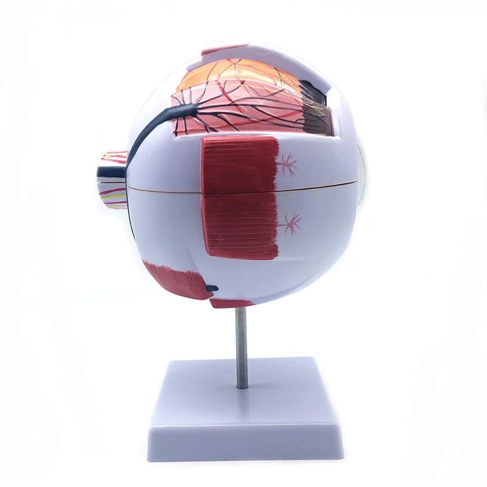
6 Times Giant Eye Model
6 Times giant eye model on base for use in patient education or anatomical study. the model display the anatomical structure of human eyeball, such as the three layers of outer membrane (outer membrane, media and intima) of eyeball wall and the main refractive bodies, lens and vitreous body filled inside. The model includes a labeled diagram and is about 9.8x6.2 x 6.2 inches (H x W x D , where H is height, the vertical distance from the lowest to highest point
Model:HSBM-186 Human Eye model
Send Inquiry
Product Description
6 Times Giant Eye Model
The best Medical education model, made of high quality PVC, Durable on transportioan and can be used very long time , 2 years warranty
Features of 6 Times Giant Eye Model:
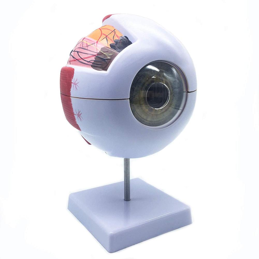
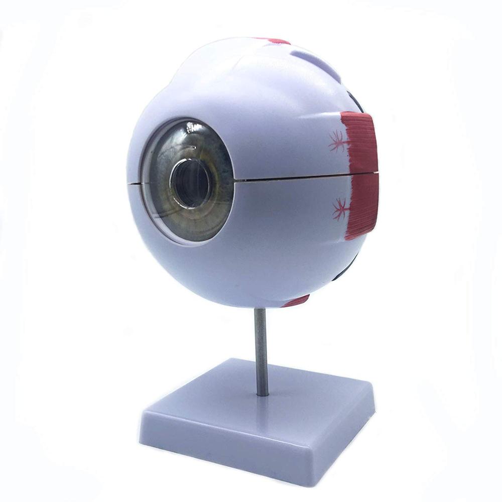
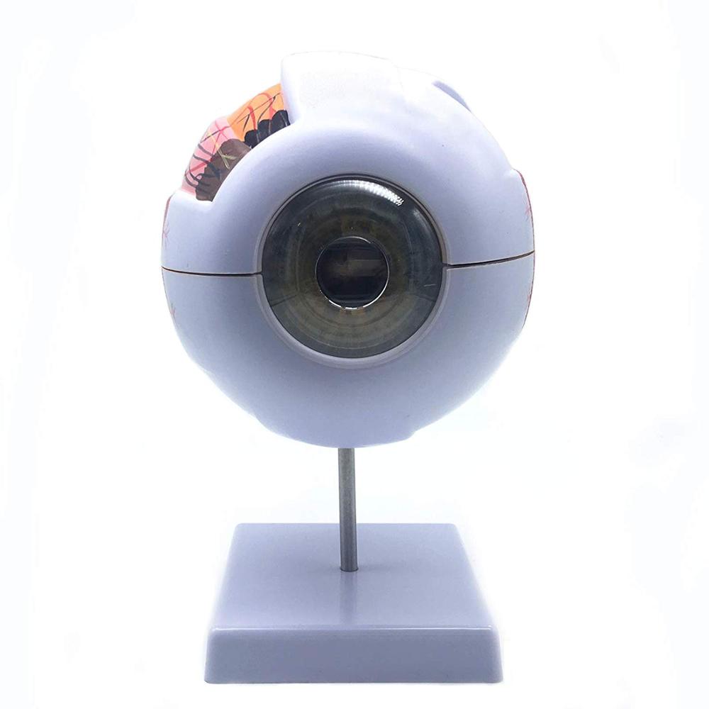
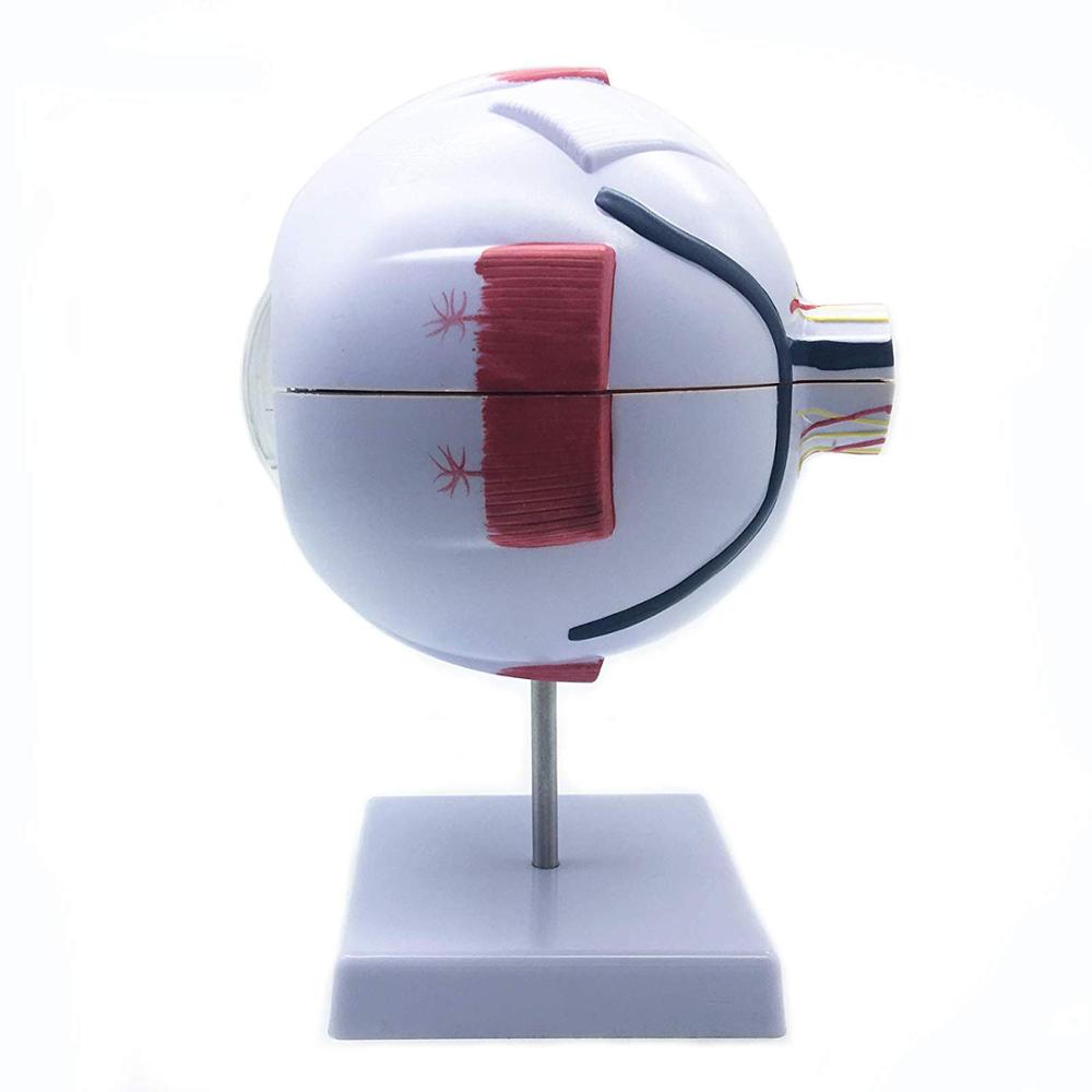
6X Enlarged Human Eye Model on base for use in patient education or anatomical study. the model display the anatomical structure of human eyeball, such as the three layers of outer membrane (outer membrane, media and intima) of eyeball wall and the main refractive bodies, lens and vitreous body filled inside. The model includes a labeled diagram and is about 9.8x6.2 x 6.2 inches (H x W x D , where H is height, the vertical distance from the lowest to highest point; W is width, the horizontal distance from left to right; D is depth, the horizontal distance from front to back). Anatomical models are typically used as educational aids in medical and scientific classrooms and office settings.
Product description of 6 Times Giant Eye Model
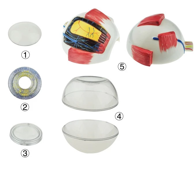
Enlarged. About 6 times of life size. Comes with a horizon section of the eyeball wall. Sclerotic membrane, choroids membrane, lens and vitreous humor are removable. The blood supply and the origins of the muscles control the eyeball movement are also shown. On stand with base. 6 parts.
The different parts of the eyeball model are detachable to show the following structures.
1. Tunica external: showing cornea and sclera with attachments of ocular muscles and optic nerve.
2. Tunica media: showing the iris, the ciliary body and the chorioid.
3. Tunica internal is retina.
4. Refraction media: showing the lens and the vitreous body. 6 times enlarged. On stand.
Carton size: 37*37*49cm 12pcs/carton
G/W.: 12kgs/carton
N/W.: 10kgs/carton